
Int J App Pharm, Vol 18, Issue 1, 2026, 306-313Original Article
BAOBAB EXTRACT WITH ZINC OXIDE NANOPARTICLES FOR TARGETED TREATMENT FOR LIVER CANCER
AYMN YASEEN SHARAF ZEEBAREE1, ABEER MANSOUR ABDEL RASOOL2* , MAES MK. ALKHYATT3
1Pharmaceutical Chemistry Department, College of Pharmacy, Ninevah University-Ninevah City-Iraq. 2,3Pharmacology and Toxicology Department, College of Pharmacy, Ninevah University-Iraq
*Corresponding author: Abeer Mansour Abdel Rasool; *Email: abeer.mansour@uoninevah.edu.iq
Received: 09 Jun 2025, Revised and Accepted: 24 Nov 2025
ABSTRACT
Objective: Hepatocellular carcinoma (HCC) is presently one of the most common and most deadly malignancies in the world, and it has a poor prognosis and a limited number of treatment strategies. Conventional treatment usually has serious side effects and resistance. The current study was designed to compare the effectiveness of Adansonia digitata L. (baobab) fruit extract as a green biosynthetic agent, zinc oxide nanoparticles (ZnNPs@BF) and to determines potential anticancer activity on liver cancer.
Methods: ZnO nanoparticle synthesis was performed using the baobab extract as a natural reducing and stabilizing agent. The phytochemical content of baobab, the process involved in nanoparticle fabrication, and the physicochemical characteristics of ZnNPs@BF were established. An overview of the in vitro studies on cytotoxicity was provided, with particular focus on the comparison of liver cancer cells to normal ones.
Results: The synthesized ZnNPs@BF by the green approach showed excellent properties with a homogeneous hexagonal structure and an average crystallite size of 33 nm using FESM and XRD techniques. These unique properties have been exploited to assess the ZnNPs@BF anti-cancer activity. The anti-cancer test revealed that ZnNPshave potent dose-dependent cytotoxicity against HCAM liver cancer cells with IC50 of 1.45 μg/ml and showed no cytotoxicity with normal HBL-100 cells. These findings highlight that ZnNPs@BF can play a crucial role as a promising, effective, and selective anti-cancer nanomaterial with other biomedical potential applications.
Conclusion: The zinc oxide nanoparticles (ZnNPs@BF) prepared from baobab extract are a safe, environmentally friendly, and effective method for delivering drugs to liver cancer cells. However, based on the obtained results, further preclinical and clinical research is needed to confirm their use and translate them into clinical applications.
Keywords: Baobab extract, Zinc oxide nanoparticles, Liver cancer, Drug delivery, Cell proliferation
© 2026 The Authors. Published by Innovare Academic Sciences Pvt Ltd. This is an open access article under the CC BY license (https://creativecommons.org/licenses/by/4.0/)
DOI: https://dx.doi.org/10.22159/ijap.2026v18i1.55835 Journal homepage: https://innovareacademics.in/journals/index.php/ijap
INTRODUCTION
The advance of potential nanoparticles with uncommon physical, chemical, and biological properties has made nanotechnology transform most of the other scientific corrections, particularly medicine [1]. Cancer of the liver is one of the greatest common and lethal malignancies in the world, with therapeutic interventions and a poor prognosis. The grouping of current therapy strategies, which include chemotherapy and targeted therapeutics, tends to have severe adverse properties and the ability to cause drug resistance. Therefore, it is strongly required to identify novel, benign, and effective therapeutic agents [2].
Hepatocellular Carcinoma is one of the most common tumors that has been found mostly as a killer caused by cancer [3]. This is usually quite difficult with its high occurrence, poor prognosis and cannot be significantly detected early enough To some extent, this is indeed diagnosed at such an advanced stage of the disease where it becomes impossible to perform surgical resections or even carry out liver transplant This notwithstanding, there are some advantages as associated with conventional treatment methods such as chemotherapy, radiotherapy, and targeted therapies, but they are normally fraught with high-side effects and high levels of resistance leading to the overall low survival rate in patients. In addition, recurrence of liver cancer following the first-line therapies is among the most topical issues, which points to the necessity to improve and less toxic methods of treatment [4].
Latest investigations exhibit an optimistic view of nanoparticles, particularly the ones based on bio plant extracts, as chemotherapeutic agents. These nanoparticles can deliver effectively to a specific location with a low toxicity towards healthy tissue, further enhancing the clinical performance without minimizing the side effects [5].
Adansonia digitata. L, often called the Tree of Life, and which goes under the name of baobab fruits, has bioactive components such as polyphenols, flavonoids, and antioxidants that are said to be behind the properties of the fruits. The phytochemicals, however, make baobab a trained candidate in the production of nanoparticles [6]. Specifically, the extract of baobab fruit has been investigated in relation to its capacity to serve as a natural reducing and capping agent during the synthesis of nanoparticles. The addition of the baobab extracts to zinc oxide (ZnO) nanoparticles also increases their application as drug delivery agents, as it strengthens the capacity of the nanoparticle to enter the cancer cells [7].
Of all these, flavonoids, tannins, and polyphenols displayed significant effects in the modulation of cell signaling, reduction of oxidative stress, one of the major players involved in the development and progression of cancer disease, and inhibition of proliferation of cancerous cells [8]. In fact, the antioxidant effect was clarified in the n-butanol isolation of the baobab fruit pulp by showing an action similar to the standards, including ascorbic acid. In this respect, antioxidants against free oxygen radicals have a critical role in combating oxidant stress, which commonly results in cancer. Besides, it is reported that its baobab fruit pulp extract in combination with ZnO nanoparticles has demonstrated amplified anti-cancer origins with greater effects on Hep-G2 adscititious liver cancer. All these combinations would maximize vivo bioavailability and cellular internalization potential of the nanoparticle, a reliable vehicle for targeting liver cancer selectively. Baobab extract incorporated ZnO nanoparticle is promising not only as an anticancer agent; it is also a hepatoprotective and drug delivery platform, which appears as a new development in the treatment of liver cancer [9].
The synthesis of ZnNPs has been used to create ZnNPs@BF, through which the healing properties of the plant are integrated with the working properties of the nanoparticle invention in cancer treatment. The phytoconstituents improved the cancer cell specificity of ZnNPs in activating and regulating important signaling pathways, in addition to the reduction of the side effects of the conventional therapy. ZnNPs are among such molecules that have become one of the most powerful particles in drug delivery and antimicrobial, and cancer therapeutics due to their distinctive properties. The use of plant extracts in the biosynthesis of the ZnNP has become an eco-friendly process, replacing conventional chemical syntheses. In this method, hazardous chemicals are minimized, and the behavior of natural reducing and stabilizing agents existing in the extracts of plants is employed [10].
The current research aims to support, describe, and investigate the cytotoxicity of zinc oxide nanoparticles incorporated with baobab extract against liver cancerous cells. The biocompatibility to the determination of ZnNPs@BF on normal cell lines demonstrates a greener and promising therapeutic alternative in the treatment of liver cancer. Additional modifications or details ought to be drawn to my attention.
MATERIALS AND METHODS
Utilized materials
The salt of zinc (II) chloride [purity: 98%; CAS No. 7646-85-7; molecular weight: 136.30 g/mol] was acquired from Sigma-Aldrich Company. The fresh fruits of Adansonia digitata L. (baobab) were purchased at the local herbal market in Nineveh City, Nineveh Governorate, Iraq. The botanical identity of the plant material was ascertained by an expert in Pharmacognosy, College of Pharmacy, Ninevah University, and a voucher specimen (Voucher No. NU-AD-2024-15) was deposited in the Herbarium of Ninevah University as a reference for future use. Human liver cancer cells (HCAM) and normal human breast epithelial cells (HBL-100).
Baobao fruit extract preparation [BFE]
In the present experiment, 1 g of BF powder was added to 100 ml of distilled water in a 250 ml conical flask. This was subsequently incubated at 70 °C for 30 min, after which it was left to cool to room temperature. Following the process, it was filtered on Whatman No. 1 filter paper to remove solid residues. This was solved into a clear form, in which the filtrate was then kept at 4 °C to undergo other tests [11].
ZnNPs@BF fabrication
Different concentrations of the solution were prepared by dissolving 0.5 g, 1g, 1.5 g, and 2 g of ZnCl2.5H2O in different, individually clean beakers. Next, 15 ml of the previously prepared (1µg/100 ml) crude BF extract was dropped into all the solutions prepared using zinc chloride. Blend the brew constantly for a couple of hours to achieve uniformity. 1 ml of 1 M sodium hydroxide [NaOH] was added to each mixture, which was placed on a magnetic stirrer at ambient temperature and left overnight not heating. Once in the stable state, the mixtures were centrifuged at 15,000 rpm/10 min. The resulting ZnNPs@BF was rinsed with double-distilled water and dried at room temperature for 2 d [12].
ZnNPs@BF diagnosis
The preparation and characterization of ZnNPs in the aqueous extract of Baobab fruit were examined in a double-beam UVvis spectrophotometer (i1900 UV/Vis Spectrophotometer, SHIMADZU, USA, Ninevah University) at a wavelength of 200 to 750 nm to record the characteristic peak to prove the ZnNPs@BF formation. To determine the functional groups and the phytoconstituents that may contribute to the reduction and capping of the ZnNPs@BF, the FT-IR was performed using RS Affinity, SHIMADZU, USA, Ninevah University, across the range of 4000-400 1/cm. Each of these field emission scanning microscope (ZEISS-SEM, UK), high resolution-transmission electron microscope (JSM-1400, Japan), energy dispersive X-ray (EDX-PLANUS), and X-ray diffractometer(Philips PW1730X nomatoPRO) was used to examine surface morphology, elemental ratio, shape, and size of green synthesized ZnNPs@BF [12].
Cell lines
The present study was conducted to evaluate the effect of Nano baobab on the liver cell line HCAM normal human HBL100 cell lines (HBL100). Cells were grown in RPMI-1640, which was supplemented with 10% Fetal bovine serum, 100 units/ml penicillin, and 100 μg/ml streptomycin. The passaging of the cells was enacted through trypsin-EDTA. The cells were reseeded at fifty percent confluence twice per week and incubated at 37C and five percent carbon dioxide [13].
Cytotoxic assays
The MTT cell viability assay was performed on 96-well plates. According to the trial protocol, cell lines were put at a density of 1*104 cells per well. The different concentrations of nano baobab (0.5, 1, 1.5, 2, 2.5) µg/ml were added to the well after replacing the old medium with fresh culture medium. Starting 24 h after, and based on the creation of a confluent monolayer after 72 h, cells were treated with various concentrations of nano baobab and reported cytotoxicity on the normal cell and liver cancer by using an equation (1) below [14]:

Determination of maximum inhibitory concentration (IC50)
Non-linear regression analysis using GraphPad Prism version 8 was employed to calculate the IC-values. Results are represented by the mean+/-SD of three independent experiments (n=3) remained applied after 72 h on the liver cancer cell line and healthy cell lines [15].
Statistical analysis
The Statistical Software for Social Sciences (SPSS version 24) was used for the statistical analyses and data preparation, and the mean and standard error were determined. P-values were calculated by ANOVA and the Duncan test post hoc to determine significant differences between and within groups. Results were performed and presented as means±SEM using the statistical software GraphPad Prism version 8. P values* denote significant when p0.05, and **denote highly significant when p0.01 [16].
RESULTS AND DISCUSSION
Optical properties of ZnNPs@BF
The synthesized nanoparticles of ZnO were systematically considered and evaluated for their anticancer effects. Initially, the synthesized NPs were tested using UV-Vis spectroscopy. Fig. 1 shows the UV-visible spectrum of the BF extract and ZnO nanoparticles, which is expected of the BF extract. The BF has shown absorbance peaks at 329.24 nm, with ZnNPs@BF biosynthesized showing absorbance of 360.11 nm. The blue shift of the absorbance is strong evidence of the size decrease of bulk molecules to the nano size. This can be attributed to the transition of materials whereby they start at a lower energy level and, through the capture of energy, they move intoa higher energy level, i. e., the red shift that is associated with quantum confinement effects and the size-dependent optics of nanoparticles. The present study also observed that, depending on the structural and electronic properties of the samples, as well as any change in bandgap energy on the nanoscale of ZnNPs@BF. The findings may indicate the suitability of ZnNPs@BF in being use in anticancer therapy, whether it will be in drug delivery, photothermal therapy, or production of reactive oxygen species. Such optical properties are also relevant to the functionality of ZnNPs@BF, the use of which can be utilized in tissue imaging or in treatment, especially. Anticancer us therapy.
FT-IR study of ZnNPs@BF
The FT-IR analysis was employed to identify simple functional groups in designed molecules. In the following case, The FT-IR spectra were performed with BF extract and the formatted ZnNPs@BF to research the alterations of the chemical groups that might take place in the two samples. The FT-IR spectra shown in fig. 2 were recorded both on the untouched composition of BF and on the disintegration of this substance with a solution of zinc chloride, and observed that the wide and narrow peaks of the BF extract and ZnNPs are provided. At 3273.75 1/cm, the present is that of N-H stretching of amine, whereas the weak band at 2920.12 1/cmis attributed to the H-C-H symmetric stretching in alkanes. Capsaicin, an alkaloid, is characterized by an N-H stretch. The band at 1017.11 1/cm is ascribed to C-O stretching in fruit extract. Simultaneously, the band at 3249.91 1/cm in nanoparticles would be the O-H stretch of hydrogen-bonded Phenols and alcohols. The band 2930.17 1/cm 2849.98 is assigned to the C-H symmetric stretch of the alkane’s formula. The significant increase in peak intensity at the 1020.03 1/cm position is attributed to the C-O stretch of the ether structure. The synthesis of zinc nanoparticles may also involve secondary metabolites, mostly as proteins binding with zinc ions. Lastly, the presence of an intense band at 544 1/cm is seen as an indication of the typical absorption of Zn-O site binding, which linkage occurs with hydro change groups present in BF bio molecules. These observed values are quite close to the recorded reports in the same domain in the past. To elucidate the formation process and stability of the ZnNPs@BF, the current study used the FT-IR spectrum, which indicates the kind of interaction and functional groups that were involved in the formation process and stabilization of the ZnNPs@BF. The observed shifts of the peaks are evidence of effective coordination of the phytochemicals of the extract of baobab to the ions of zinc, which confirms the synthesis of the nanoparticles, the way in which the biomolecules of the BF extract are bonding the ions of zinc to the synthesis of the nanoparticles by embracing functional groups. The formation of ZnNPs is accompanied by shifting peaks; a chemistry where addition to other things listed in the paper, a signature is the Zn-O bond, signifier of their genesis [17, 18].
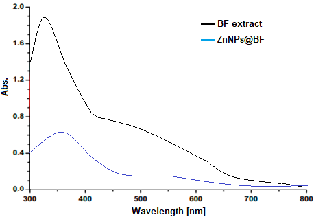
Fig. 1: UV-Vis diagnosis of BF extract and ZNPs@BF
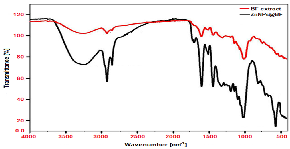
Fig. 2: FT-IR diagnosis of BF extract and ZnNPs@BF
XRD study of ZnNPs@BF
X-ray diffraction (XRD) is a well-established diagnostic tool to determine the structural attributes and crystalline size of nanoparticles synthesized. The XRD analysis of the green obtained zinc nanoparticles is presented in fig. 3. The X-ray diffraction analyses showed outstandingly intense peaks that appeared at certain degrees of 2 degrees (2 theta) 30.58, 33.29, 37.26, 45.41, 57.31, 60.99, 66.78, and 69.01. These diffraction peak values correspond to the lattice planes (100, 002, 101, 102, 110, 103, 112, and 201). Based on the obtained XRD pattern, it was determined that all the peaks were in agreement with the standard JCPDS file [card no. 76-0704. This ensures that the syntheses ZnNPs@BF have a hexagonal crystalline structure. The XRD analysis can provide excellent information concerning the crystalline character and the structural diagnosis of the synthesized zinc nanoparticles. The agreement of the diffraction peaks with the reference data confirms the validity of the crystal structure and proves that the ZnNPs@BF synthesis was successful and can take the shape of a hexagon.
The size of ZnOs crystallite, D, of ZnNPs@BF was determined using the formula, of Debye Scherrer [D = k 01540/2 0.94 or D = K/degrees lambda] where the variables are, i. e. the diameter of the ZnO crystalline equal to the value of D; the wavelength of an X-ray source (0.15406 in XRD); β is full width half maximum of a certain diffraction peak; the value of Scherer (0.94) and The size of the result obtained was 33 nm. The acquired outcome confirms the effectiveness of the green synthesis methodology established to produce uniformly crystalline ZnNPs@BF, and, thus, the route can be used in properties of interest and could be used in other fields of research as well [17].
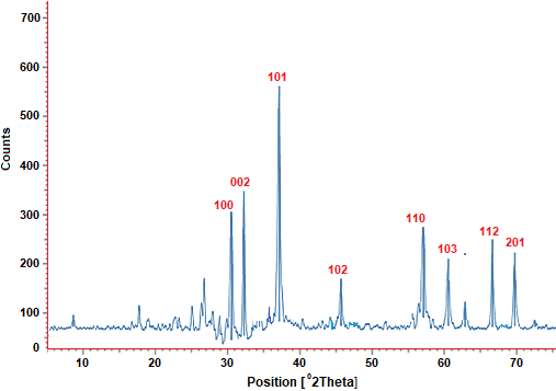
Fig. 3: XRD analysis of ZnNPs@BF
Morphology formation of ZnNPs@BF
This had been done by carrying out FESEM, HR-TEM, and EDX investigations to reveal more properties of the sustainably designed ZnNPs@BF. A good regular crystalline character of the nano-structural homogeneities with more or less hexagonal morphologies of ZnNPs was demonstrated by the FESEM micrograph (fig. 3a). Such a structure was sustained and observed deep-probed through HR-TEM (fig. 4b) image analysis that indicated that there was an excellent arrangement of ZnNPs in the same form as it occurred in the FESEM diagnosis image. Besides, the observed measurement was comparable with the results of XDR analysis, which ensured that the green NPs were prepared satisfactorily. Besides these diagnoses, the energy-dispersive X-ray spectroscopy (EDS) analysis spectrum was used on the sustainable fabricated ZnNPs@BF (fig. 3c). The spectrum recorded indicated an excellent strong main peak of the zinc element at optimal expected position of 0.9-1.21 keV, which is in accordance with the nominal position of metallic Zn (fig. 4c). The peaks of high signals of carbon [C] and oxygen [O] elements were observed in a range of 0.01-0.21 and 0.4-0.72keV respectively. The interaction between two components essentially is characterized by the existence of the linkage of phytochemicals with the surface of ZnNP. XRSEM, HR-TEM, and EDX have revealed that the ZnNPs@BF possess a very homogeneous structure, particularly consistent size, and constant elemental structure. This demonstrated that the green synthesis technique is efficient to generate quality nanoparticles of controlled properties [18].
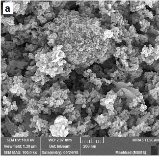
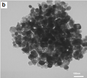
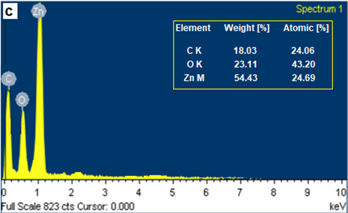
Fig. 4: (a) FESEM analysis, (b) HR-TEM image diagnosis, and (c) EDX assay of ZnNPs@BF
Evaluation effect of Nano baobab on the liver cell line HCAM
Evaluation effect of Nano baobab on liver cell line HCAM after 48 h
The current study employed the MTT assay to evaluate the cytotoxicity of this substance on a human liver cell line over 48 h. The results are shown in table 1. Statistical determination in three replications showed that the difference was significant compared to the control. Based on the results obtained, it was identified that, as the concentration of nano baobab continues to increase on HCAM cells after 72 h, a decline in the percentage of cell survival rate is observed (63.47 %, 40.77 %, 33.54 %, 25.93% and 22.10%), with these values and significant changes in zero concentration (before the addition of Nano baobab). The potential use of nano baobab as an anti-proliferative agent is shown by the feature that growth of cancer cells is inhibited, as demonstrated in several studies and illustrated in table 1 and fig. 5. Nonetheless, the cytotoxic effects exhibited demonstrate that its dose needs to be optimized to prevent off-target effects. Such a compromise is a vital step to the highest possible effectiveness of therapy and impairment reduction. However, additional studies (long exposure and use on a variety of cell lines) are necessary to get a clear picture of its safety and efficacy [19]. Aqueous extracts of baobab leaves were found to have an anticancer effect on gastric and osteosarcoma cancer cell lines, when it was found that specific metabolites play a role in its effectiveness, and some of these metabolites identified include catechin and rutin [20].
Table 1: Cytotoxic effect of nano baobab on liver cell line after 48 h
| Concentration µg/ml | 0 | 0.5 | 1 | 1.5 | 2 | 2.5 |
| Mean | 97.10 d | 63.47 c | 40.77 d | 33.54 d | 25.93 a | 22.10 a |
| SEM | 1.993 | 4.944 | 3.804 | 3.441 | 4.913 | 2.027 |
| P-Value | 0.000*** | |||||
| Post Hoc Duncan's Test | ||||||
| 0 vs. 0.5 | **0.0049 | |||||
| 0 vs. 1 | ***0.0004 | |||||
| 0 vs. 1.5 | ***0.0004 | |||||
| 0 Post Hoc Duncan's Test vs. 2 | ****0.000 | |||||
| 0 vs. 2.5 | ****0.000 | |||||
| 0.5 vs 1 | * 0.012 | |||||
| 0.5 vs. 1.5 | **0.0039 | |||||
| 0.5 vs. 2 | 0.000* | |||||
| 0.5 vs. 2.5 | 0.003** | |||||
| 1 vs. 1.5 | 0.3050ns | |||||
| 1 vs. 2 | 0.450ns | |||||
| 1 vs. 2.5 | 0.093ns | |||||
| 1.5 vs. 2 | 0.9150ns | |||||
| 2 vs. 2.5 | ns0.2896 | |||||
Analysis by one-way ANOVA, Post–Hoc Duncan′s test.
|
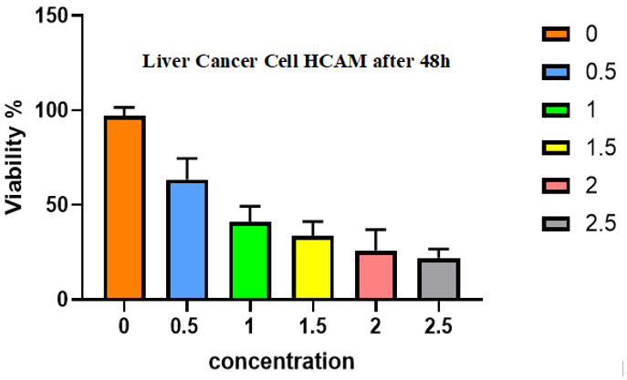
Fig. 5: Cytotoxic effect of nano baobab on liver cell line after 48h. Error bars indicate SD values
Evaluation effect of nano baobab on liver cell line after 72 h
In the present study, the MTT assay was employed to evaluate the cytotoxic effect of nano baobab on the human liver cell line over a 72-hour incubation period. The results are shown in table 2. A statistical determination was triplicated, which showed a significant difference from the control points. Based on the acquired findings, it was revealed that as the concentration of nano baobab increased on HCAM cells at 72 h, the gain in cell survival rate percentage reduced (45.67%, 17.54%, 13.87%, 12.75%, and 11.40%), which was significantly varying to zero concentration (before adding the nano baobab) as it was reflected in table 2 and fig. 6.
Evaluation impact of nano baobab between 48 h and 72 h
By comparing 48 h and 72 h, the current study not only reveals that there is no significant difference up to 48 h before treatment, but also notices differences between all concentration levels at about 48 h and 72 h, as shown in fig. 7 by paired test. Nano baobab cytotoxicity effect on a liver cell line after 72 h can be interpreted within the broader context of nanomaterials causing cytotoxicity on liver cells, namely: the HepG2 cell line. Although no studies exist on Nano baobab, it is possible to derive knowledge using similar nanomaterials in their interaction with the liver cells [21].
Table 2: Cytotoxic effect of nano baobab on liver cell line after 72 h
| Concentration µg/ml | 0 | 0.5 | 1 | 1.5 | 2 | 2.5 |
| Mean | 93.77 d | 45.67 c | 17.54 b | 13.87 b | 12.75 a | 11.40 a |
| SEM | 2.346 | 2.399 | 1.257 | 0.5432 | 0.3711 | 0.6784 |
| P-Value | 0.000*** | |||||
| Post Hoc Duncan's Test | ||||||
| 0 vs. 0.5 | **0.01 | |||||
| 0 vs. 1 | ****0.000 | |||||
| 0 vs. 1.5 | ****0.000 | |||||
| 0 vs. 2 | ****0.001 | |||||
| 0 vs. 2.5 | ****0.00 | |||||
| 0.5 vs 1 | **0.032 | |||||
| 0.5 vs. 1.5 | 0.0012** | |||||
| 0.5 vs. 2 | 0.010** | |||||
| 0.5 vs. 2.5 | 0.0187** | |||||
| 1 vs. 1.5 | 0.213ns | |||||
| 1 vs. 2 | 0.0198* | |||||
| 1 vs. 2.5 | 0.298ns | |||||
| 1.5vs. 2 | ns0.9150 | |||||
| 1.5 vs. 2.5 | ns0.2896 | |||||
| 2 vs. 2.5 | ns0.9597 | |||||
|
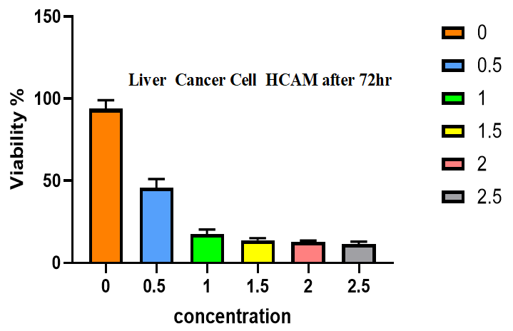
Fig. 6: Cytotoxic effect of nano baobab on liver cell line after 72h. Error bars indicate SD values
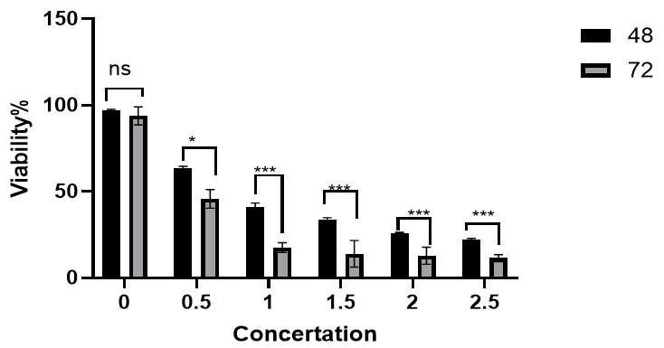
Fig. 7: Cytotoxic effect of nano baobab on liver cell line after 48 and 72 h (ns: non-significant)*=less than 0.05, **less than 0.001 *** less than 0.0001. Error bars indicate SD values
Evaluation Impact of nano baobab on normal cell line (HBL-100) after 72 h
Cytotoxic effect on the normal cell line was non-significant between all concentrations and before adding, as shown in fig. 8. Cytotoxicity of nano-baobab to a normal cell line (which is HBL 100) was measured after a 72 h incubation period, and no significant cytotoxicity levels were produced among all concentrations in comparison to the baseline level before the insertion of the nanoparticles. Consequently, implies that nano-baobab does not render harm to the viability of the HBL-100 cells that are derived from human breast milk and have transformed cell-like characteristics, as indicated by Gaffney in 1981 [21]. The result supports the study of nanoparticles, in which the toxicity of nanoparticles can be out of control, depending on their composition and the types of cells they encounter [22]. Nano baobab based on Adansonia digitata L. The idea of investigating the possible use of nano baobab as an anticancer agent because its phytochemical contents [22]. The Lack of cytotoxicity on the HBL-100 cells implies that Nano baobab can be nontoxic to the normal cells and hence safe to be used in the proposed therapy [23].
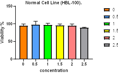
Fig. 8: Cytotoxic effect of nano baobab on normal cells after 72h. Error bars indicate SD values
Determination of Half-maximal inhibitory concentration [IC50] of nano baobab
The IC50 of ZnNPs@BF against HCAM liver cancer cells was 1.45±0.12 μg/ml, whereas it did not show an IC50 on HBL-100 normal cells, even at the highest concentrations tested on fig. 9a and b, on the anticancer efficacy of nanoparticles and even those obtained in nature, and especially on Nano of Adansonia digitata L, which towers above the rest of the anti-cancer agents to attack only liver cancers without damaging the normal cells. The IC50 can be viewed as one of these vital parameters, as it indicates the concentration that has led to the suppression of 50 % viability in the cells. As far as the themes of the nanoparticles derived from baobab are concerned, they have been found to exhibit a significant cytotoxic effect on cancerous cells, particularly those related to the liver, including HepG2, but not on normal cell lines, hence leading to a selective anticancer activity. Baobab extracts have shown great anti-cancer activity on HepG2 cancer cells of the liver, and the IC50 value is very significant, as it shows that it has a very good inhibitory effect on cancer cell proliferation [24]. Other studies involving the use of nanoparticles like gold nanoparticles and the iron oxide nanoparticles have been demonstrated to cause similar cytotoxicity, with most effectiveness being rendered to the liver cancer cells, with just very insignificant toxicity being exhibited on normal cells [25].
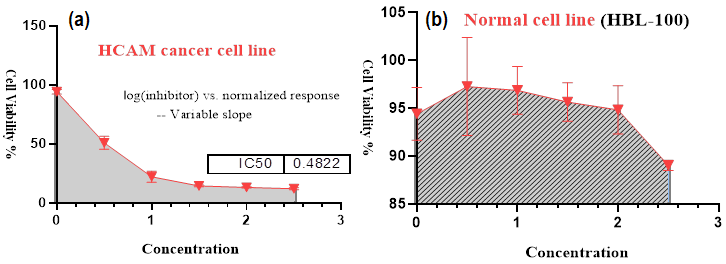
Fig. 9: (a) Half-maximal inhibitory concentration [IC50] of nano baobab in liver cancer cells, (b) Half-maximal inhibitory concentration IC50 of nano baobab in normal cancer cells. (The IC50 values were calculated by non-linear regression analysis (GraphPad Prism 8)
CONCLUSION
Finally, the optical capabilities of ZnNPs@BF play a key role in increasing their potential as anticancer agents. Such properties allow the application to perform more complex diagnosis, imaging, and emergent therapies, including photothermal and photodynamic therapies. This knowledge of the relationship of these nanoparticles with light enables the playing of optimization so as to ideally and effectively target cancer as an effective form of therapeutic cancer treatment and cure, which involves precision of diagnosis and treatment. Consequently, the study of the optical properties of ZnNPs@BF is extremely important in the evolution of this nanomaterial in the management of cancer.
ACKNOWLEDGMENT
Our research group would like to thank the University of Nineveh and the College of Pharmacy for their assistance, guidance and support in completing this work.
FUNDING
Nil
AUTHORS CONTRIBUTIONS
Aymn Yaseen Sharaf Zeebaree: Conceptualization, Writing-original draft. Maes MK. Alkhyatt; Conceptualization, Writing-original draft. Abeer Mansour Abdel Rasool; Conceptualization, Writing-review and editing.
CONFLICT OF INTERESTS
The authors declare that there is no conflict of interest.
REFERENCES
Bhutadiya VL, Mistry KN. A review on bioactive phytochemicals and its mechanism on cancer treatment and prevention by targeting multiple cellular signaling pathways. Int J Pharm Pharm Sci. 2021;13(12):15-9. doi: 10.22159/ijpps.2021v13i12.43031.
Begum SA, Rani SJ, Banu A, Pavani A, Yeruva V. Statistics of cancer, 2020 in Indian states: a review on the report from national cancer registry programme. Asian J Pharm Clin Res. 2021;14(6):36-42. doi: 10.22159/ajpcr.2021.v14i6.41616.
Dutta S, Deb N, Pattnaik AK, Besra SE. Apoptosis-inducing potential of Lawsonia alba Lam. leaves on hepatocellular carcinoma (HepG2) cells along with its antioxidant property. Int J Pharm Pharm Sci. 2016;8(9):156-62. doi: 10.22159/ijpps.2016.v8i9.12815.
Allaire M, Goumard C, Lim C, Le Cleach A, Wagner M, Scatton O. New frontiers in liver resection for hepatocellular carcinoma. JHEP Rep. 2020;2(4):100134. doi: 10.1016/j.jhepr.2020.100134, PMID 32695968.
Gokul M, Umarani G, Esakki A. Green synthesis and characterization of isolated flavonoid mediated copper nanoparticles by using Thespesia populnea leaf extract and its evaluation of antioxidant and anticancer activity. Int J Chem Res. 2022;6(1):15-32. doi: 10.22159/ijcr.2022v6i1.197.
Adesina JA, Zhu J. A review of the geographical distribution, indigenous benefits and conservation of African baobab (Adansonia digitata L.) tree in sub-Saharan Africa. Preprints. 2022 May 23;1. doi: 10.20944/preprints202205.0287.
Neamah SA, Albukhaty S, Falih IQ, Dewir YH, Mahood HB. Biosynthesis of zinc oxide nanoparticles using Capparis spinosa L. fruit extract: characterization, biocompatibility and antioxidant activity. Appl Sci. 2023;13(11):6604. doi: 10.3390/app13116604.
Kopustinskiene DM, Jakstas V, Savickas A, Bernatoniene J. Flavonoids as anticancer agents. Nutrients. 2020;12(2):457. doi: 10.3390/nu12020457, PMID 32059369.
Vinha AF, Costa AS, Pimentel FB, Espirito Santo L, Sousa C, Freitas M. Bioactive compounds and scavenging capacity of Adansonia digitata L. (Baobab fruit) pulp extracts against ROS and RNS of physiological relevance. Appl Sci. 2024;14(8):3408. doi: 10.3390/app14083408.
Menazea AA, Ismail AM, Samy A. Novel green synthesis of zinc oxide nanoparticles using orange waste and its thermal and antibacterial activity. J Inorg Organomet Polym Mater. 2021;31(11):4250-9. doi: 10.1007/s10904-021-02074-2.
Ijoma KI, Ajiwe VI. Phytochemical screening of Dialium indum leaf extract (Velvet tamarind). Int J Phytopharmacy. 2017;7(1):6-13. doi: 10.7439/ijpp.v7i1.3942.
Ji X. A perspective of ZnCl₂ electrolytes: the physical and electrochemical properties. eScience. 2021;1(2):99–107. doi: 10.1016/j.esci.2021.09.003.
Hoseini Alfatemi SM, Fallah F, Armin S, Hafizi M, Karimi A, Kalanaky S. Evaluation of blood and liver cytotoxicity and apoptosis-necrosis induced by nanochelating-based silver nanoparticles in mouse model. Iran J Pharm Res. 2020;19(2):207-18. doi: 10.22037/IJPR.2020.1101026, PMID 33224226.
Chung DM, Kim JH, Kim JK. Evaluation of MTT and trypan blue assays for radiation-induced cell viability test in HepG2 cells. Int J Radiat Res. 2015;13(4):331. doi: 10.7508/ijrr.2015.04.006.
Rasool MA, Qader KO, Mustafa NW. Anticancer promise of Loranthus europaeus extract: impact on the AMJ13 breast cancer cell line. Health Biotechnol Biopharma (HBB). 2025;9(2):117–34. doi: 10.5281/zenodo.13802725.
Chinnasamy S, Ramchandran M, Selvam M, Nanjundan P. Alternative energy exploitation of agricultural biomass using SPSS Statistics. J Appl Chem Phys. 2023;2(4):1-9. doi: 10.46632/jacp/2/4/1.[17]S.
Goutam P, Yadav AK, Das AJ. Coriander extract mediated green synthesis of zinc oxide nanoparticles and their structural, optical and antibacterial properties. NanoScience Technol. 2017;3(1):249-52. doi: 10.30799/jnst.079.17030205.
Shehu A, Joshua OO, Uduma UA. Unveiling the potential of a tri-seed synthesis approach: synthesis of zinc nanoparticles with enhanced antimicrobial properties. Discov Chem. 2025;2(1):1-19. doi: 10.1007/s44214-025-00055-3.
Kadam SD, Kondawar MS. Evaluation of in vitro anticancer activity and quantitation of active ingredient of Adansonia digitata L. fruit. Int J Pharm Sci Drug Res. 2019;11(6):358–69. doi: 10.25004/IJPSDR.2019.110613.
Chouhan MK, Roy TK, Patil D, Bhatkal A, Kasajima I, Hegde S. Cytotoxicity assessment and LC-MS profiling of Adansonia digitata on human gastric and osteosarcoma cancer cell lines. Food Hum. 2024;2:100270. doi: 10.1016/j.foohum.2024.100270.
Gaffney EV. A cell line (HBL-100) established from human breast milk. Cell Tissue Res. 1982;227(3):563-8. doi: 10.1007/BF00204786, PMID 6891286.
Pola S, Konatala A. Evaluation of toxicity of nanoparticles using cell lines. In: Siddhardha B, Dyavaiah M, Kasinathan K, editors. Model organisms to study biological activities and toxicity of nanoparticles. Singapore: Springer Singapore; 2020. p. 297-315. doi: 10.1007/978-981-15-1702-0_15.
Chouhan MK, Roy TK, Patil D, Bhatkal A, Kasajima I, Hegde S. Cytotoxicity assessment and LC-MS profiling of Adansonia digitata on human gastric and osteosarcoma cancer cell lines. Food Hum. 2024 May;2:100270. doi: 10.1016/j.foohum.2024.100270.
Kadam SD, Kondawar MS. Evaluation of in vitro anticancer activity and quantitation of active ingredient of Adansonia digitata L. fruit. Int J Pharm Sci Drug Res. 2019;11(6):358–69. doi: 10.25004/IJPSDR.2019.110613.
Mikaeili Ghezeljeh S, Salehzadeh A, Ataei E Jaliseh S. Iron oxide nanoparticles coated with glucose and conjugated with Safranal (Fe3O4@Glu-Safranal NPs), inducing apoptosis in liver cancer cell line (HepG2). BMC Chem. 2024;18(1):33. doi: 10.1186/s13065-024-01142-1, PMID 38360669.