
Int J Curr Pharm Res, Vol 18, Issue 2, 31-37Original Article
EVALUATION OF ANTI-DEPRESSANT ACTIVITY OF ETHANOLIC EXTRACT OF PIPER METHYSTICUM LEAVES IN MICE
DEEPALI THAKUR*, AJEET PAL SINGH, AMAR PAL SINGH
Department of Pharmacology, St. Soldier Institute of Pharmacy, Lidhran Campus, Behind NIT (R. E. C.), Jalandhar –Amritsar by pass, NH-1, Jalandhar-144011, Punjab, India
*Corresponding author: Deepali Thakur; *Email: deepali.thakur010@gmail.com
Received: 14 Nov 2025, Revised and Accepted: 01 Jan 2026
ABSTRACT
Objective: The aim of this study to evaluate the anti-depressant activity of ethanolic extract of Piper methysticum leaves in mice.
Methods: The animals were grouped into four groups, each comprising 6 mice. One of the group as control, receiving normal saline water and second group as standard, receiving fluoxetine, while the remaining two groups were administered the test drug (300 mg/kg and 600 mg/kg). All drug administrations were conducted orally 1 h prior to the acute study test procedure and daily for a 14 d period for the chronic study.
Results: The administration of Piper methysticum doses resulted in a notable reduction in immobility duration during the Tail Suspension Test. In the Actophotometer assessment, mice exhibited a statistically significant increase in motor activity compared to the control group. The findings indicated that the ethanolic extract of Piper methysticum leaves at a dose of 600 mg/kg led to a significant increase in reduced glutathione and catalase activity. Simultaneously, there was a decrease in plasma corticosterone, lipid peroxidation, plasma nitrite, and protein concentration levels.
Conclusion: The antidepressant efficacy of Piper methysticum at a dosage of 600 mg/kg in mice is comparable to that of fluoxetine.
Keywords: Antidepressant, Fluoxetine, Piper methysticum, Oxidative stress, Major depressive disorder.
© 2026 The Authors. Published by Innovare Academic Sciences Pvt Ltd. This is an open access article under the CC BY license (https://creativecommons.org/licenses/by/4.0/)
DOI: https://dx.doi.org/10.22159/ijcpr.2026v18i2.8017 Journal homepage: https://innovareacademics.in/journals/index.php/ijcpr
INTRODUCTION
Depression, sometimes referred to as major depressive disorder (MDD), is a complicated condition with a wide range of symptoms. These include a chronically depressed mood, a loss of interest and enjoyment in normally enjoyable activities, a decrease in energy, difficulties thinking and making decisions, changes in appetite and sleep patterns, psychomotor issues, and thoughts of suicide [1]. It drastically lowers a person's quality of life by impairing their social, professional, and educational functioning. Additionally, MDD is linked to a higher risk of death and suicide [2]. It has been continuously shown that dysregulated hypothalamic-pituitary-adrenal (HPA) axis activity at different hierarchical levels is a hallmark of major depressive disorder (MDD). In particular, corticotrophin-releasing hormone (CRH) levels in cerebral fluid are elevated in MDD patients [3]. In basic care settings, behavioural health issues are often overlooked. It is noteworthy that depression, which affects at least 8% of primary care patients, shows that only 50% of depressed people are usually recognised by primary care research [4]. 2004 Around 350 million people worldwide suffer from depression, according to the World Health Organisation, which also revealed that depression is the third largest cause of disease worldwide, after diarrhoeal illness and lower respiratory infections, and before ischaemic heart disease and HIV/AIDS. According to projections, depression is expected to move up to the top of this list by 2030 [5, 6]. It is thought that pharmacological treatments for severe depressive disorders alter the central monoaminergic systems, particularly affecting noradrenergic synaptic neurotransmissions and serotonin (5-HT). A significant portion of patients, around one-third of them, show only partial or no reaction to the recommended treatment, even though selective serotonin reuptake inhibitors and noradrenaline reuptake inhibitors successfully treat the bulk of depressive episodes. Therefore, the goal of current research is to find novel antidepressants that are more effective [7]. Natural therapeutic agents having great promise for treating a variety of illnesses, including anxiety and depression, include medicinal plants and their extracts. Many herbs have been used in folk medicine for many centuries to treat depression. Using well-established methods to evaluate antidepressant-like effects in animal animals, a variety of plant extracts and natural products have been investigated as possible antidepressant drugs [8]. Originating in the South Pacific, kava (Piper methysticum) is a plant whose roots have long been used in traditional medicine for cold-water, non-alcoholic extractions. The goal of this therapy is to treat a number of illnesses, such as menstruation problems, anxiety, tension, pain, and muscle spasms. The six main lipophilic kavalactones in kava are responsible for its therapeutic effectiveness; the most powerful anxiolytic effects are shown by kawain and dihydrokawain. It has been suggested that the limbic regions of the brain are where kavalactone acts primarily. Kavalactones have a variety of neurobiological actions that contribute to their anxiolytic effects, but they primarily involve modulating gamma-aminobutyric acid (GABA) receptors by blocking voltage-gated sodium ion channels, reducing excitatory neurotransmitter release by blocking calcium ion channels, and improving ligand binding to GABA type A receptors [9, 10]. However, its possible antidepressant-like effect and the possible modification of monoaminergic connection to this activity have not yet been investigated. As previously mentioned, Piper methysticum has been recognised for its anxiolytic and neuroprotective qualities [10]. Therefore, the current study was designed to investigate the antidepressant-like effects of Piper methysticum in mice that were not under stress and mice who were under stress from acute immobilisation. The study also seeks to clarify the possible underlying processes linked to this behaviour that has been seen.
MATERIALS AND METHODS
Animals
We bought Swiss mice of both sexes (12–24 w old, 20–30 g in weight) from Lovely Professional University Animal House in Jalandhar. Separate cages of six animals each were kept in a polycarbonate cage measuring 29×22×14 cm. The animals were kept in a laboratory setting with a 12 h cycle of light and dark. The behavioural tests were conducted between 9:00 and 17:00 h after the animals had been acclimated for at least fourteen days. In accordance with the guidelines of the Committee for the Purpose of Control and Supervision of Experimental Animals (CPCSEA), Ministry of Environment and Forests, Government of India (Reg. No. 2011/PO/Re/S/18/CPCSEA, with registration date of 01/05/2018), the experimental protocol was approved by the Institutional Animals Ethics Committee (IAEC) via letter number IAEC/SSIP/2023/PR-032, dated May 2023, for the use and care of experimental animals.
Drugs and chemicals
Piper methysticum was obtained from Kshipra Biotech Pvt. ltd. We bought fluoxetine from the pharmacies. Starting on the eighth day, fluoxetine was administered orally at a dosage of 10 mg/kg. The above concentrations were created by dissolving the drugs individually in double-distilled water. Every chemical and biological reagent utilised in this investigation was of analytical quality and was made just before use.
Experimental design
Swiss mice (either sex) were weighed and divided into 4 groups, each consisting of 6 animals. They were treated once per day for 14 d as follows:
Group 1: control group, normal saline 0.9% NaCl.
Group 2: mice treated with fluoxetine (10 mg/kg, orally) suspended in distilled water.
Group 3: mice treated with Piper methysticum ethanolic leaves extract (PMELE)-300 mg/kg suspended in distilled water.
Group 4: mice treated with Piper methysticum ethanolic leaves extract (PMELE)-600 mg/kg suspended in distilled water.
Acute oral toxicity
Low, medium, and high doses were chosen for therapy in an acute trial for an ethanolic extract of Piper methysticum conducted in accordance with OECD recommendations No. 423. Approach: Four groups of six mice each were created from the overnight fasting mice. Using a gavage, the ethanolic leaf extract was administered orally in dosages of 5, 50, 300, and 1000. The animals were monitored for tremors, convulsions, salivation, diarrhoea, lethargy, sleep, and coma for the first two and twenty-four hours after the extract was administered. They were also watched for toxic symptoms and mortality for up to fourteen days. The survivor mice were restored and used again for experiments after 14 d of acute oral toxicity [11]. Three mice were discovered dead after four to twenty-four hours of treatment with 1000 mg/kg body weight. Consequently, doses of 300 and 600 mg/kg body weight were chosen based on the aforementioned finding.
Behavioural model
Tail suspension test
A frequently used technique for assessing novel antidepressant medications is the tail suspension test. Drugs like imipramine and fluoxetine, which are therapeutically successful in treating human depression, may reverse immobility, which is a sign of helplessness. In this experimental paradigm, the length of immobility during a certain time period is used as the measure of depression. The lengthening of the immobility period is a sign of mental depression. On the other hand, a shorter length of immobility indicates a state of mind free from depression [7]. Thirty minutes before testing, groups of six animals are given either the vehicle or the test compounds orally. Adhesive tape is used to hang the mice on the edge of a shelf 58 cm above a tabletop for the test, about 1 cm from the tip of their tails. Five minutes are allotted for recording the length of immobility. When mice hang passively and without moving for at least a minute, they are said to be immobile.
Locomotor activity
The locomotors activity can be an index of wakefulness (alertness) of mental activity. The locomotors activity can be easily measured using an actophotometer. Activated the Actophotometer to verify the proper functioning of all photo cells to ensure precise recording. Subsequently, individual mice were positioned within the activity cage for a duration of 10 min, during which the activity scores were documented for each animal up to the specified time limit. Finally, the observed motor activity was assessed and juxtaposed with the control or standard.
Biochemical estimation
Collection of blood samples
Blood (0.3 ml) was extracted from the tail vein of every mouse group on the fifteenth day. A cooled centrifuge (Paramount Scientific Works, Ambala Cantt, India) was used to separate the plasma from blood samples at 2500 rpm for 10 min. This plasma was then utilised to estimate the levels of corticosterone.
Preparation of brain homogenate
Animal brains were dissected and washed in ice-cold saline on the fifteenth day. The cerebellum was thrown away. 10% (w/v) tissue homogenates were made in 0.1 M phosphate buffer (pH 7.4) for biochemical assessments. After centrifuging the homogenates for 15 min at 10,000 g, aliquots of the supernatants were extracted and utilised for the catalase test, nitrite, reduced glutathione, and lipid peroxidation estimation.
Estimation of oxidative stress
Estimation of plasma corticosterone levels
The approach of Bartos and Pesez (1979) was used to quantitatively estimate the amount of corticosterone in the blood plasma. After immersing the tubes in cold water for five minutes, 0.50 ml of 0.10 N sodium hydroxide was added to 1.0 ml of the sample in ethanol along with 0.50 ml of a 0.10% solution of p-nitroso-N, N-dimethylaniline in ethanol. After being sealed with cotton-wool, the tubes were left in a dark place at 0 °C for five hours. Five millilitres of a 1.0% aqueous potassium ferricyanide solution, two millilitres of pH 9.8 buffer, and five millilitres of a 0.10% phenol in ethanol solution were added to the aforesaid solution. For ten minutes, the tubes were maintained at 20±2 °C in a water bath. A UV-visible spectrophotometer (UV 3200 UV-VIS Spectrophotometer, Somajiguda, Hyderabad) was used to read the solution at 650 nm.
Estimation of plasma nitrite levels
The technique was used to quantify plasma nitrite. A 5% aqueous solution of m-phosphoric acid (1 part) and 0.1% w/v N-(1-Naphthyl) ethylene diaminedihydrochloride (1 part) were combined with 1%w/v sulphanilamide and allowed to sit at 0 °C for 60 min. After mixing 0.5 ml of plasma with 0.5 ml of the aforementioned combination, the mixture was left at room temperature in the dark for 10 min. A UV-visible spectrophotometer (Varian Cary 5000 UV-VIS-NIR Spectrophotometer, Netherlands CHRIST) was used to measure the absorbance at 546 nm.
Estimation of protein concentration
A total protein kit (Siemens, Siemens Ltd., Vadodara, Gujarat) and a semi-automatic autoanalyzer (Chem 5 plus-V2 semi-autoanalyzer; Erba Mannheim, Germany) were used to assess the total protein content in brain homogenate.
Procedure
The Biuret technique was used to measure the total protein content at a wavelength of 546 nm. The steps used were those outlined in the brochure that came with the kit (Henry and Winkelman, 1974).
Its components are as follows: 1) Reagent 1 (Biuret Reagent): potassium iodide (30 mmol/l), sodium hydroxide (3.8 mol/l), potassium sodium tartrate (0.1 mol/l), and cupric sulphate (33 mmol/l).
2) Surfactant (20g/l) is Reagent 1A (Surfactant).
3) BSA (60 g/l) is the standard (total protein 6 g/dl).
Reconstitution of the reagents: Let them come to room temperature. To one bottle of Reagent 1, add 41 ml of distilled water. Next, add the contents of one bottle of Reagent 1A. Gently stir to prevent foaming. Close firmly.
Before use, the sample and reconstituted reagent were allowed to come to room temperature.
This kit was utilised with the general system characteristics listed below:
Type of reaction: Final destination
546 nm is the wavelength (530–570 nm)
20 min of incubation, RT
Volume of sample: 10 µl
Volume of reagent: 1.0 ml
Reagent configuration with: At room temperature, incubate the reagent blank for 20 min. Combine and read.
Lipid peroxidation estimation
The technique was used to assess the amount of malondialdehyde, a lipid peroxidation indicator, in the form of thiobarbituric acid-reactive compounds. In short, two hours were spent incubating 0.5 ml of post-mitochondrial supernatant and 0.5 ml of Tris-HCl at 37 °C. Following incubation, 1 ml of 10% trichloroacetic acid was added, and the mixture was centrifuged for 10 min at 1,000 rpm. After adding 1 ml of 0.67% thiobarbituric acid to 1 ml of supernatant, the tubes were placed in boiling water for ten minutes. Following cooling, 1 ml of double-distilled water was added, and a UV-visible spectrophotometer (Varian Cary 5000 UV-VIS-NIR Spectrophotometer, Netherlands CHRIST) was used to detect absorbance at 532 nm. Using an extinction coefficient of 1.56×10⁵ M-1 cm-1, thiobarbituric acid-reactive compounds were measured and expressed as nanomoles of malondialdehyde per milligramme of protein.
Reduced glutathione estimation
We used the Jollow et al. (1974) technique to measure reduced glutathione. In short, 1.0 ml of sulfosalicylic acid (4%), mixed with 1.0 ml of post-mitochondrial supernatant (10%), precipitated the mixture. After being stored at 4 °C for at least an hour, the samples were centrifuged for 15 min at 4 °C at 1,200 rpm. A total of 3.0 ml of the test mixture included 0.1 ml of supernatant, 2.7 ml of phosphate buffer (0.1M, pH 7.4), and 0.2 ml of 5,5 dithiobis-(2-nitrobenzoic acid) (Ellman's reagent, 0.1 mmol, pH 8.0). At 412 nm, a UV-visible spectrophotometer (Varian Cary 5000 UV-VIS-NIR Spectrophotometer, Netherlands CHRIST) was used to measure the yellow colour that had evolved. The molar extinction value of 1.36×10⁴ M−1 cm−1 was used to compute GSH levels, which were then reported as micromole per milligramme of protein.
Estimation of catalase activity
The Claiborne (1985) technique was used to measure catalase activity. In short, the test mixture was composed of 1.0 ml hydrogen peroxide (0.019 M), 1.95 ml phosphate buffer (0.05 M, pH 7.0), and 0.05 ml post-mitochondrial supernatant (10%), totalling 3.0 ml. A Varian Cary 5000 UV-VIS-NIR Spectrophotometer (Netherlands CHRIST) was used to measure changes in absorbance at 240 nm. The millimolar extinction coefficient of H2O2 (0.07 mmol) was used to measure catalase activity, which was then represented as micromoles of H2O2 degraded per minute per milligramme of protein.
RESULTS
Tail suspension test
In this test statistically significant reduction in immobility was observed across all groups, except for the control group (256±3.828 seconds) (P<0.05). While a decrease in the immobility period was evident in the Piper methysticum 300 mg/kg group (62±2.744 seconds), Piper methysticum 600 mg/kg group (34±2.666 seconds), and fluoxetine group (19.4±1.626 seconds). However, in a group receiving Piper methysticum at a dose of 600 mg/kg, the reduction in immobility was significantly similar as compared to the group receiving fluoxetine.
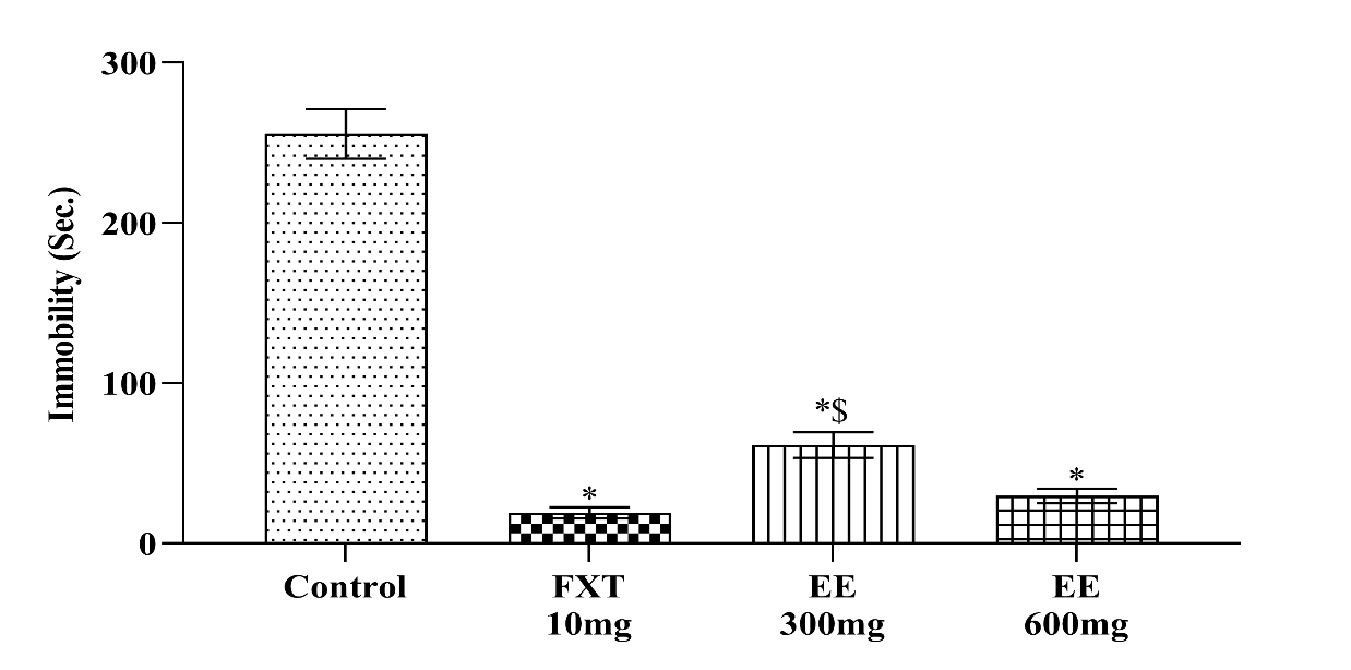
Fig. 1: Effects of ethanolic extract of leaves of Piper methysticum and Fluoxetine on duration of immobility in the tail suspension test
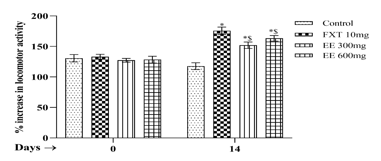
Fig. 2: Effects of ethanolic extract of leaves of piper methysticum and Fluoxetine on locomotor activity
Locomotor activity
The reduction in locomotor activity was notably pronounced following immobilization in comparison to mice subjected to controlled treatment. Piper methysticum and Fluoxetine, employed in the current investigation, demonstrated a significant impact on the spontaneous locomotor activity of mice in contrast to their respective control cohorts.
Value are expressed as mean±SEM (One-way ANOVA followed by Tukey’s test). In fig. 2, the mice of Piper methysticum ethanolic leaves extract (300 and 600 mg/kg/p. o) group showed significantly difference in locomotors activity as compared to control group. Treatment with Fluoxetine (10 mg/kg p. o) also showed maximum significant change as compared to control group.
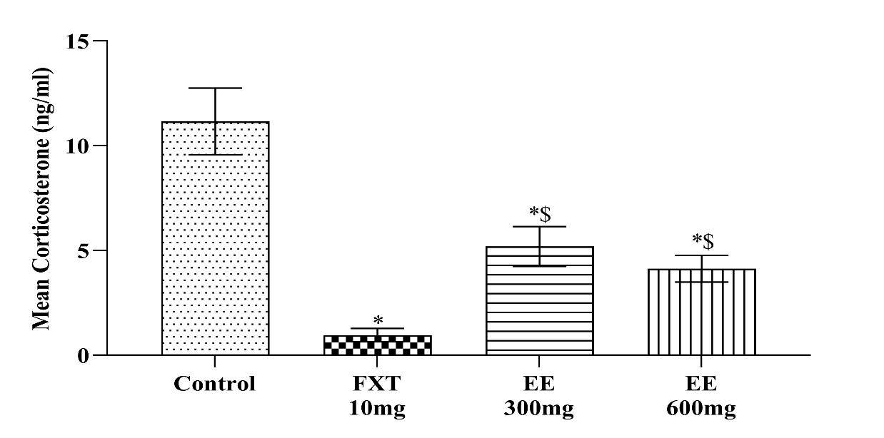
Fig. 3: Effects of ethanolic extract of leaves of piper methysticum and fluoxetine on plasma corticosterone level
Normally in depressive mice, corticosterone level is high. As shown in fig. 3, after administration of ethanolic extract of leaves of Piper methysticum as 300 and 600 mg/kg and standard Fluoxetine (10 mg/kg) have decreased the corticosterone level. Alcoholic extract of leaves of Piper methysticum 600 mg/kg produced a greater activity, which is comparable to that of standard Fluoxetine (10 mg/kg).
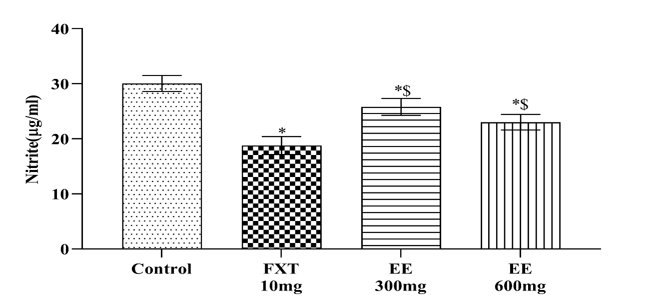
Fig. 4: Effects of ethanolic extract of leaves of piper methysticum and fluoxetine on plasma nitrite level
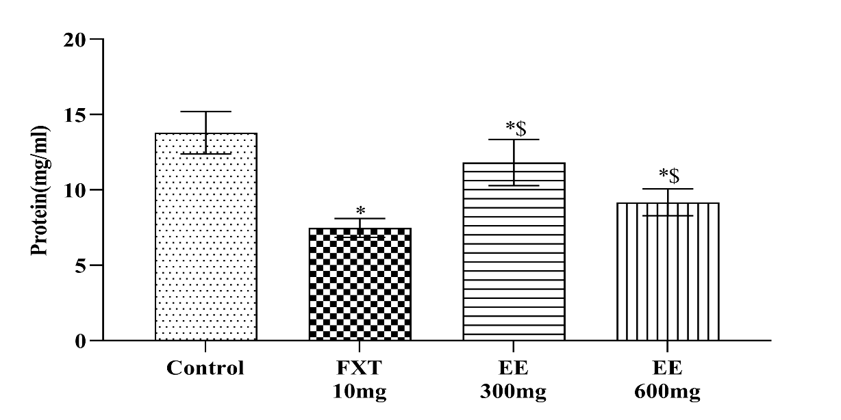
Fig. 5: Effects of ethanolic extract of leaves of piper methysticum and fluoxetine on plasma protein level
Estimation of plasma nitrite level
In stressed animal, a decrease in cerebral blood is intimately related to increase in nitrite levels. After administration of ethanolic extract of leaves of Piper methysticum as 300 and 600 mg/kg and standard Fluoxetine (10 mg/kg) have decreased the plasma nitrite level. As shown in fig. 4, alcoholic extract of leaves of Piper methysticum 600 mg/kg produced a greater anti-depressant activity which is comparable to that of standard Fluoxetine (10 mg/kg).
Estimation of protein concentration level
In depressive and aged animals, both albumin and total protein value increased dramatically. After administration of ethanolic extract of leaves of Piper methysticum as 300 and 600 mg/kg and standard Fluoxetine (10 mg/kg) have decreased the plasma protein concentration level. In fig. 5, alcoholic extract of leaves of Piper methysticum 600 mg/kg produced a greater activity which is comparable to that of standard Fluoxetine (10 mg/kg).
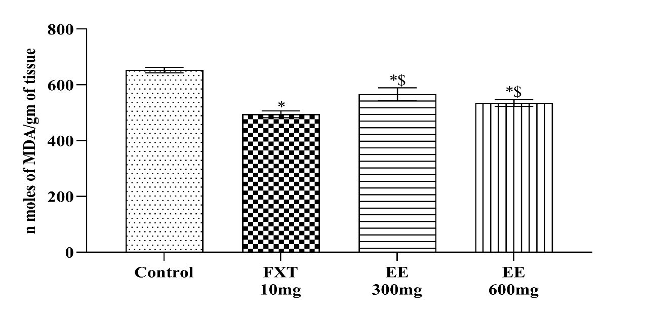
Fig. 6: Effects of ethanolic extract of leaves of piper methysticum and fluoxetine on lipid peroxidation level
Estimation of lipid peroxidation level
Lipid peroxidation level greater in depressive animals. As shown in fig. 6, after administration of ethanolic extract of leaves of Piper methysticum as 300 and 600 mg/kg and standard Fluoxetine (10 mg/kg) have decreased the lipid peroxidation level. Alcoholic extract of leaves of Piper methysticum 600 mg/kg produced a greater activity which is comparable to that of standard Fluoxetine (10 mg/kg).
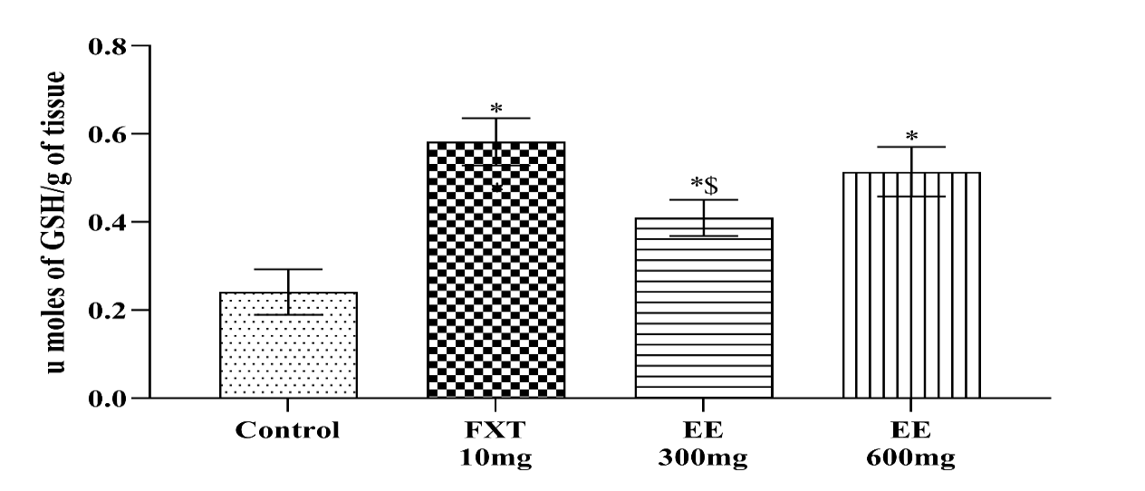
Fig. 7: Effects of ethanolic extract of leaves of piper methysticum and fluoxetine on reduced GSH level
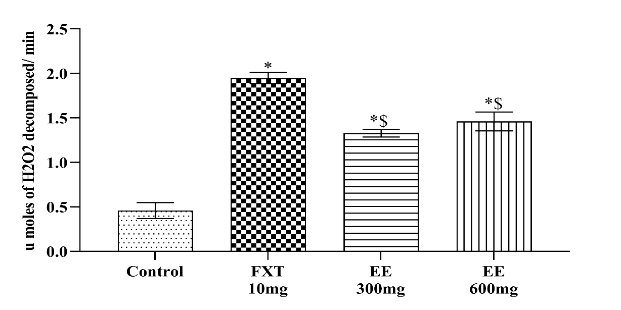
Fig. 8: Effects of ethanolic extract of leaves of piper methysticum and fluoxetine on catalase activity
Estimation of reduced glutathione activity
A significant decline in GSH level is observed in depressive animals. As shown in fig. 7, after administration of ethanolic extract of leaves of Piper methysticum as 300 and 600 mg/kg and standard Fluoxetine (10 mg/kg) have increased the reduced GSH level. Alcoholic extract of leaves of Piper methysticum 600 mg/kg produced a greater activity, which is comparable to that of standard Fluoxetine (10 mg/kg).
Estimation of catalase activity
In depressed mice, reactive oxygen species are produced, which set off a chain of events that leads to neuronal death. The brain has several defences, but is nevertheless susceptible to oxidative damage because of its high levels of ROS and declining levels of antioxidants like catalase, GSH, and SOD and others. So in depressive mice there is significantly decrease the activity of catalase in the brain. Fig. 8 shown that after administration of ethanolic extract of leaves of Piper methysticum as 300 and 600 mg/kg and standard Fluoxetine (10 mg/kg) have restored the reduced activity of catalase in brain. Alcoholic extract of leaves of Piper methysticum 600 mg/kg produced a greater activity which is comparable to that of standard Fluoxetine (10 mg/kg).
DISCUSSION
Community depression is high and associated to a lot of illness. Therefore, these difficulties must be addressed and effective remedies found [12]. Pharmaceutical treatments for depression have limitations, thus alternative therapies are needed. Herbal depression treatments may be effective. Over the last decade, herbal intervention research has advanced, highlighting the growing interest in its therapeutic potential. Piper methysticum is one of several naturally occurring components and plant parts that have pharmacological action.
Ethanolic extracts from Piper methysticum were given orally to Swiss mice at 300 and 600 mg/kg to test their antidepressant qualities. Traditional medicine uses the herb to relieve tension. Our research found several phytoconstituents in Piper methysticum leaf alcohol that had antidepressant properties. Our investigation supports the mythical antidepressant properties of Piper methysticum leaves.
TST and actophotometer were used to determine antidepressant-like action. These mouse behavioural despair models assess immobility decrease to predict antidepressant effectiveness. This immobility, called behavioural despair in animals, may mimic human sadness [7]. The Tail Suspension Test (TST) and Actophotometer showed a dose-dependent reduction in immobility time for Piper methysticum ethanolic extracts compared to the control group. Immobility time decreased significantly in mice treated with Fluoxetine (10 mg/kg).
Numerous studies link stress to increased free radical production [13]. Free radicals promote corticosterone release due to hypothalamic-pituitary-adrenal axis overactivation [14, 15]. Stress-induced free radicals cause metabolic imbalances and harmful effects, according to several studies (Olivenza, 2000). Oxidative stress is an imbalance in oxidation-reduction processes that reduces the antioxidant defence system's ability to combat excess oxygen-derived molecules. This causes oxygen poisoning and associated side consequences. In depressed people, oxidative stress is elevated [16]. Increased malondialdehyde (MDA) levels in stressed mice' brains indicate oxidative damage [17]. Lipid peroxidation exacerbates the depletion of intracellular endogenous antioxidant glutathione, a primary defence against oxidative damage, due to the brain's elevated oxygen tension, reduced antioxidant capacity, and abundant supply of oxidisable substrates [18]. At all dosages, Piper methysticum reduced plasma corticosterone, plasma protein, plasma nitrite, lipid peroxidation, and stress-induced brain glutathione and catalase depletion. These data suggest Piper methysticum may reduce stress-related physiological abnormalities. Alcoholic Piper methysticum leaf extract 600 mg/kg has antidepressant efficacy equivalent to Fluoxetine. These difficulties may lead to a new age of therapeutic treatment employing plant-derived principles that can benefit humans.
In stressed mice, Piper methysticum 600 mg/kg orally had the strongest antidepressant effect. Thus, this dose was chosen to study its antidepressant processes.
CONCLUSION
In conclusion, the administration of Piper methysticum at a dosage of 600 mg/kg has exhibited antidepressant efficacy in mice that is comparable to fluoxetine. The findings from the current investigation provide a basis for future exploration into the mechanisms underlying the antidepressant action of Piper methysticum. Consequently, additional research endeavors in this direction are imperative to elucidate the specific mechanisms through which Piper methysticum operates in the context of depression.
ACKNOWLEDGMENT
It’s our privilege to express the profound sense of gratitude and cordial thanks to our respected chairman Mr. Anil Chopra and Vice Chairperson Ms. Sangeeta Chopra, St. Soldier Educational Society, Jalandhar for providing the necessary facilities to complete this research work.
AUTHORS CONTRIBUTIONS
All the authors have contributed equally.
CONFLICT OF INTERESTS
Declared none
REFERENCES
Ghoneim MM, O’Hara MW. Depression and postoperative complications: an overview. BMC Surg. 2016 Feb 2;16:5. doi: 10.1186/s12893-016-0120-y.
Chaabna K, Cheema S, Abraham A, Maisonneuve P, Lowenfels AB, Mamtani R. The state of population health research performance in the Middle East and North Africa: a meta research study. Syst Rev. 2021;10(1):1. doi: 10.1186/s13643-020-01552-x, PMID 33388080.
Glynn LM, Davis EP, Sandman CA. New insights into the role of perinatal HPA-axis dysregulation in postpartum depression. Neuropeptides. 2013;47(6):363-70. doi: 10.1016/j.npep.2013.10.007, PMID 24210135.
Jeffrey J, Do MT, Hajal N, Lin YH, Linonis R, Grossman MS. Using web-based technology to improve depression screening in primary care settings. BMJ Open Qual. 2021;10(1):e001028. doi: 10.1136/bmjoq-2020-001028, PMID 33589504.
Chuang CY, Shi YC, You HP, Lo YH, Pan TM. Antidepressant effect of GABA-rich Monascus-fermented product on forced swimming rat model. J Agric Food Chem. 2011;59(7):3027-34. doi: 10.1021/jf104239m, PMID 21375324.
Hamid HA, Ramli AN, Yusoff MM. Indole alkaloids from plants as potential leads for antidepressant drugs: a mini review. Front Pharmacol. 2017;8:96. doi: 10.3389/fphar.2017.00096, PMID 28293192.
Dhingra D, Chhillar R. Antidepressant-like activity of ellagic acid in unstressed and acute immobilization-induced stressed mice. Pharmacol Rep. 2012;64(4):796-807. doi: 10.1016/S1734-1140(12)70875-7, PMID 23087132.
Moragrega IS, Rios JL. Medicinal plants in the treatment of depression: evidence from preclinical studies. Planta Med. 2021;87(9):656-85. doi: 10.1055/a-1338-1011, PMID 33434941.
Savage KM, Stough CK, Byrne GJ, Scholey A, Bousman C, Murphy J. Kava for the treatment of generalised anxiety disorder (K-GAD): study protocol for a randomised controlled trial. Trials. 2015;16:493. doi: 10.1186/s13063-015-0986-5, PMID 26527536.
Dragull K, Yoshida WY, Tang CS. Reactive oxygen species and reactive nitrogen species: relevance to cyto(neuro)toxic events and neurologic disorders. J Agric Food Chem. 2012;59(3):207-13. doi: 10.1021/jf203345p.
Khan MA, Tiwari SB, Gupta H, Noor H. Evaluation of anxiolytic and antidepressant potential of hydro-alcoholic leaves extract of Azadirachta indica in albino rats. Pharmacologyonline. 2020;3:207-13.
Yeung KS, Hernandez M, Mao JJ, Haviland I, Gubili J. Herbal medicine for depression and anxiety: a systematic review with assessment of potential psycho-oncologic relevance. Phytother Res. 2018;32(5):865-91. doi: 10.1002/ptr.6033, PMID 29464801.
Liu J, Wang X, Shigenaga MK, Yeo HC, Mori A, Ames BN. Immobilization stress causes oxidative damage to lipid protein and DNA in the brain of rats. FASEB J. 1996;10(13):1532-8. doi: 10.1096/fasebj.10.13.8940299, PMID 8940299.
Qin B, Zhou X, Michael KD, Liu Y, Whittington C, Cohen D. Psychotherapy for depression in children and adolescents: study protocol for a systematic review and network meta-analysis. BMJ Open. 2015;5(2):e005918. doi: 10.1136/bmjopen-2014-005918, PMID 25681311.
Liu L, Liu C, Wang Y, Wang P, Li Y, Li B. Herbal medicine for anxiety, depression and insomnia. Curr Neuropharmacol. 2015;13(4):481-93. doi: 10.2174/1570159X1304150831122734, PMID 26412068.
Shoeb A, Chowta MN, Pallempati G, Rai A, Singh A. Evaluation of antidepressant activity of vanillin in mice. Indian J Pharmacol. 2013;45(2):141-4. doi: 10.4103/0253-7613.108292, PMID 23716889.
Soares RB, Dinis Oliveira RJ, Oliveira NG. An updated review on the psychoactive, toxic and anticancer properties of kava. J Clin Med. 2022;11(14):4039. doi: 10.3390/jcm11144039, PMID 35887801.
Metodiewa D, Koska C. Reactive oxygen species and reactive nitrogen species: relevance to cyto(neuro)toxic events and neurologic disorders. Neurotox Res. 2000;1(3):197-233. doi: 10.1007/BF03033290, PMID 12835102.